| MUSCLE INJURY | A very valuable use of thermography is in detecting muscle injury. It locates the area of inflammation associated with a muscle or muscle group. It can also show an area of atrophy before it becomes apparent clinically. Atrophy is seen as an area of consistent decrease in circulation when compared to the opposite side. Thermography allows us to see the specific location of strained or torn muscles. It can also help to assess the effects that have been placed on the musculo-skeletal system after extreme exertion and the extent of damage, or secondary damage, after a fall or accident. | | | 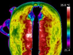 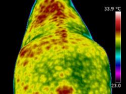 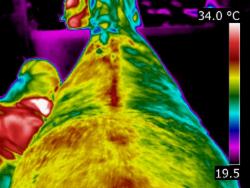
| | | When scanning initially, images of the whole horse must be assessed. If a muscle or muscle group shows a raised thermal pattern is the muscle damaged and inflamed or do these muscles simply have increased blood flow because they are working harder to compensate for injury or damage elsewhere? Only a well-trained, experienced thermographer is able to interpret the images.
When inflammation occurs in heavily muscled areas an intense thermal pattern will be evident over the affected area. If oedema and swelling occur over the strained muscle area, the surface temperature may actually become cooler due to subcutaneous fluid accumulation. Both types of images indicate an injury has occurred and careful observations are necessary to detect these differences.
| | | | |
|