| EQUINE THERMOGRAPHY ~ BENEFITSA researcher in this technology, Ram Purohit PhD, noted, ‘Thermography
may act as a ‘pain substitute’ because of its ability to detect
inflammation in the early stages before tissue damage occurs.’
Equine Thermography can be a very effective method of highlighting the location of problem areas which then require further exploration. It is a non-invasive, non-contact tool that uses the very latest infrared imaging equipment and computer software to detect minute differences in the horse’s thermal and neural condition. It is ‘real-time’, revealing changes in physiology as they are happening. The thermal pattern is the first thing that changes when things are going wrong. Equine Thermography will reveal those changes.
- Equine Thermography does not use any radiation and is therefore perfectly safe for the horse and the handler. Extensive research in human and equine fields has demonstrated that many injuries and physical conditions can be accurately detected using thermography before any physical signs and symptoms are apparent. Thermal Imaging shows the animal's physiological state by graphically mapping skin surface temperature in response to changes in blood flow, providing an objective view of the subjective feeling of pain. In healthy animals, the thermal pattern on the skin is largely symmetrical, this is because skin blood flow is controlled by the sympathetic nervous system. I am a highly trained Level 1 Thermographer and can recognise and interpret abnormal patterns.
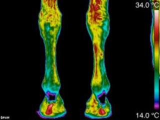
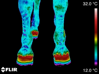
A human touch cannot identify changes in temperature of less than 2-3 degrees, however, a thermal camera can “see” and detect changes of 0.1 degree, making it easy to identify problem areas where heat is not obvious to the touch. Not only can it tell us areas of increased (abnormal) heat but importantly, areas of increased (abnormal) cool. When 'feeling' horses legs for heat it is assumed the warmer leg is abnormal, however thermal imaging can show us that the warmer leg is normal it is in fact the cooler leg that is showing us an abnormal pattern. Thermography should not be seen as a diagnostic tool but as an indicator of the 'where', it is complementary to other imaging techniques such as radiology and ultrasonography, which can then show the 'what'.
- Standard diagnostic tools that the veterinarian uses show you the damage that has been done; the actual injury, these anatomical changes are preceded by physioloical changes. Thermal Imaging is a valuable tool because it is measuring a physiological change rather than an anatomical change. Being able to spot an injury early could allow trainers to adjust training schedules and veterinarians to treat an injury before time is lost from training by a more serious injury.
‘A medical thermogram represents the surface temperature of skin, making thermography useful for the detection of inflammation,’ noted Tracy Turner DVM Associate Professor at the University Of Minnesota College Of Veterinary Medicine. ‘This ability to non-invasively assess inflammatory changes makes thermography an ideal imaging tool to aid in the diagnosis of certain lameness conditions in the horse.’ 'Thermography may have its greatest clinical application in the assessment of individual muscle injuries which are difficult to diagnose,’ noted Turner. ‘It can locate an area of inflammation associated with a muscle or muscle group and it illustrates atrophy well before it becomes apparent clinically'.
Equine thermographic imaging has proved to be a valuable tool in aiding the diagnosis and prognosis of the injured equine. By identifying the location of the injury you and your vet can make a decision on treatment and prevent further trauma. Thermography can then be used to monitor recovery.
Equine Thermography: giving your horse a voice
|
|
| | Injury monitoring Thermography assists in monitoring an injured area during rehabilitation. Thermography is generally used in conjunction with diagnostic modalities such as ultrasound. While ultrasound can follow the shape and the size of the lesion, thermography will determine when inflammation has resolved. |
| | | | |
| 
| | Injury Prevention If injury could be detected early enough progression to a more serious injury can be avoided. Dr Turner's research has shown that tendon injury can be seen up to as much as 2-3 weeks before clinical signs. |
| | | | |
| 
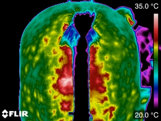
| | Clinical Entities Clinical entities that have been identified by thermography include: Horners Syndrome, Laminitis, Navicular Disease/Syndrome, hoof abscess, corns and Kissing Spines. Since thermography can assist in the detection of soft tissue inflammation, injuries occurring to tendons or ligaments may be identified. |
| | | | |
| | | | |
|
| |
|
|
|